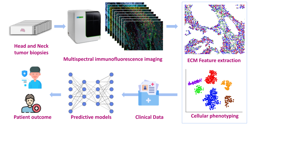
This project centers on analyzing images of head and neck cancer tissue, the 6th most prevalent cancer worldwide. In addition to standard HES histological images, we use multi-labeled fluorescence images to investigate the pathological extracellular matrix (ECM) of tumor tissue and its pivotal role in driving cancer progression and metastasis. Multi-labeled immunofluorescence imaging allows the detection of ECM components and cellular proteins in the same images, thus facilitating the study of matrix structures and their spatial distribution relative to cell phenotypes. While various cell types of the tumor microenvironment have been extensively characterized, the extracellular elements of this ecosystem remain underexplored. This project focuses on analyzing the different organizations of the ECM, aiming to correlate structural attributes with function, such as immunosuppression and cell death signaling. Our ultimate goal is to gain biologically- and clinically-relevant insights from these images and to identify prognostic and predictive biomarkers.
Clinical data are central to our investigations. We currently work on patient datasets from Unicancer-sponsored immunotherapy trials.
ECMFluo Characterizing and modeling extracellular matrix and TNF receptors in cancer from digital fluorescence microscopy
Résumé
Mots clés
- biomarkersextracellular matriximages structure analysismulti-plex immunofluorescence imagestumor microenvironment
Partenaires du projet
INS2I
Laure BLANC-FERAUD
I3S
(UMR7271) Nice
INSB
Ellen VAN OBBERGHEN-SCHILLING
IBV
(UMR7277) Nice


